What a digital X-ray really is
An X-ray is the oldest and most familiar form of medical imaging, and it remains one of the most useful tools a doctor has. When a radiographer takes an X-ray of your chest, a hand, or a tooth, a small and carefully controlled burst of energy passes through your body and lands on a detector on the other side. Dense tissues such as bone absorb more of this energy and appear bright white, while softer tissues such as lung and muscle let more of it through and look darker. The result is the familiar black and white picture that clinicians in Mauritius rely on every single day.
For most of the twentieth century these images were captured on photographic film. Today almost every hospital and imaging centre has moved to digital X-ray, and that shift has brought real benefits for patients.
How the digital version works
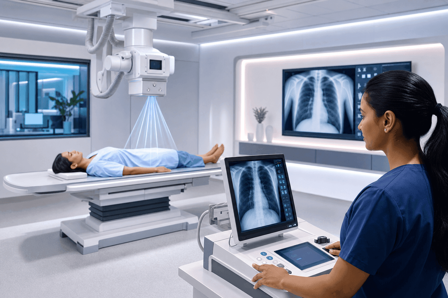
In a digital system the film is replaced by an electronic detector, similar in spirit to the sensor inside a modern camera. The moment the exposure is taken, the image appears on a computer screen. There are no chemicals to develop the picture and no waiting for it to be printed. The radiographer can check the image straight away and, if the position was not quite right, adjust it without calling you back another day.
Because the image is now a digital file, it can be brightened, magnified, and shared instantly. A radiologist can study the same picture on a high resolution monitor, and it can be sent securely to a specialist across the island or overseas within minutes.
Why digital X-ray is safer
The most important advantage is dose. Digital detectors are far more sensitive than old film, which means they need much less radiation to produce a clear image. In many cases a digital X-ray uses only a fraction of the dose that the same film study once required. Less radiation for the same quality of picture is a clear win for your long term safety.
There are other safety gains too. Because the radiographer sees the image immediately, there are fewer repeat exposures caused by a picture that turned out too dark or too light. Every avoided repeat is radiation you do not receive. Modern machines also focus the beam tightly on the area being examined, so the rest of your body is shielded and exposed as little as possible.
What a modern X-ray visit looks like
A typical X-ray takes only a few minutes. You may be asked to remove jewellery or clothing with metal, since metal blocks the beam and can hide detail. The radiographer will position the part of your body being examined against the detector and step behind a screen to take the picture. You might be asked to hold your breath for a second or two so the image is sharp. The exposure itself lasts a fraction of a second and you feel nothing at all.
If you are pregnant or think you might be, tell the radiographer before the scan. X-rays of the chest or a limb carry very little risk, but the team will always take extra care around the abdomen and will choose another test where possible.
What X-rays are good at
X-rays are quick, widely available, and inexpensive, which is why they are often the first imaging test ordered. They are excellent for looking at bones, so they are the natural choice after a fall or a suspected fracture. A chest X-ray gives a valuable first look at the lungs and heart and can point to infection, fluid, or other problems. Dentists use X-rays to see decay and the roots of teeth that are hidden from view.
They do have limits. X-rays show bone and air very well but are less good at showing soft tissue detail, which is where scans such as ultrasound, CT, and MRI take over. Your doctor chooses the test that answers the specific question about your health.
The takeaway
Digital X-ray combines a long trusted technology with modern electronics to give faster answers at a lower radiation dose. For patients in Mauritius that means a short, painless visit, an image the team can trust, and results that can be shared quickly with the doctors who need them. If you have questions about an upcoming X-ray, the radiography team is always happy to explain what to expect before you begin.
Clear imaging is the first step to an accurate diagnosis. Explore the wider Medtech health ecosystem.